Seborrheic keratosis
| Seborrheic keratosis | |
|---|---|
| Other names: Seborrheic verruca,[1] seborrhoeic keratosis,[2] barnacle,[2] senile wart,[2] seborrhoeic warts,[3] basal cell papilloma[3] | |
 | |
| Multiple seborrheic keratoses on the back of a person with cancer, known as Leser–Trélat sign. | |
| Specialty | Dermatology |
| Symptoms | Variable appearing skin growths, appears stuck on, smooth to warty[2][3] |
| Usual onset | Middle age[4] |
| Causes | Unknown[3] |
| Risk factors | Sun exposure, genetics[3] |
| Diagnostic method | Based on appearance, if unclear skin biopsy[5][3] |
| Differential diagnosis | Warts, melanoma, actinic keratosis, acanthosis nigricans[6] |
| Prevention | None[7] |
| Treatment | None, cryotherapy, laser therapy[3][2] |
| Prognosis | Harmless though persistent[2] |
| Frequency | Very common[2] |
Seborrheic keratosis are non-cancerous skin growths.[3] They have a varied appearance from flat to raised, pinpoint to a few centimeters, grey to brown to black, and smooth to warty.[2] They often appear as if stuck on.[3] They often gradually grow in size and thickness and more appear over time.[2][3] While the palms and soles are not affected, they may appear on the rest of the skin.[6] Occasionally they may itch or bleed if irritated.[2][7]
The cause is unknown.[3] Sun exposure and genetics may be a risk.[3] Rarely they occur in large numbers as a paraneoplastic syndrome.[5][7] They are not infectious.[4] They form from the cells in the outer layer of the skin.[8] Diagnosis is generally based on appearance; though, a skin biopsy may be done in unclear cases.[5][3] Other conditions that may appear similar include warts, melanoma, actinic keratosis, and acanthosis nigricans.[6]
While no treatment is generally required, some choose to have them removed by cryotherapy or laser therapy due to their appearance.[3][2] Treatment may leave an area of skin discoloration or scarring.[7] They are harmless; though, rarely resolve on their own.[2] There is no method of preventing further lesions from forming.[7] Seborrheic keratosis are common and become more so with age affecting about 30% of 40 year olds and 75% of 70 year olds.[3] The condition was first separated from warts around 1300 AD by Henri de Mondeville.[9]
Signs and symptoms
Because only the top layers of the epidermis are involved, seborrheic keratoses are often described as having a "pasted on" appearance. Some dermatologists refer to seborrheic keratoses as "seborrheic warts", because they resemble warts, but strictly speaking the term "warts" refers to lesions that are caused by human papillomavirus.[10]
-
.jpg) Seborrhoeic keratosis
Seborrhoeic keratosis -
.jpg) Seborrhoeic keratosis
Seborrhoeic keratosis -
.jpg) Seborrhoeic keratosis
Seborrhoeic keratosis
Cause
The cause of seborrheic keratosis is not known.[11]
Diagnosis
.jpg)
Visual diagnosis is made by the "stuck on" appearance, horny pearls or cysts embedded in the structure. Darkly pigmented lesions can be challenging to distinguish from nodular melanomas.[12] Furthermore, thin seborrheic keratoses on facial skin can be very difficult to differentiate from lentigo maligna even with dermatoscopy. Clinically, epidermal nevi are similar to seborrheic keratoses in appearance. Epidermal nevi are usually present at or near birth. Condylomas and warts can clinically resemble seborrheic keratoses, and dermatoscopy can be helpful. On the penis and genital skin, condylomas and seborrheic keratoses can be difficult to differentiate, even on biopsy.
A study examining over 4000 biopsied skin lesions identified as seborrheic keratoses showed 3.1% were malignancies. Two-thirds of those were squamous cell carcinoma.[13] To date, the gold standard in the diagnosis of seborrheic keratosis is represented by the histolopathologic analysis of a skin biopsy.[14]
Subtypes
Seborrheic keratoses may be divided into the following types:[15]: 767 [16][17]
| Subtype (and alternative names) | Characteristics | Image |
|---|---|---|
| Common seborrheic keratosis (basal cell papilloma, solid seborrheic keratosis) | Dull or lackluster surface.[15]: 769 | |
| Reticulated seborrheic keratosis (adenoid seborrheic keratosis) | Dull or lackluster surface, and with keratin cysts seen histologically.[15]: 769 | |
| Stucco keratosis (deratosis alba,[18] digitate seborrheic keratosis, hyperkeratotic seborrheic keratosis, serrated seborrheic keratosis, verrucous seborrheic keratosis) | Common. Dull or lackluster surface, and with church-spire-like projections of epidermal cells around collagen seen histologically.[15][19]: 637 Stucco keratoses are often light brown to off-white, and are no larger than a few millimeters in diameter. They are often found on the distal tibia, ankle, and foot.[20] | |
| Clonal seborrheic keratosis | Dull or lackluster surface, and with round, loosely packed nests of cells seen histologically.[15]: 769 | |
| Irritated seborrheic keratosis (inflamed seborrheic keratosis, basosquamous cell acanthoma) | dull or lackluster surface.[15]: 769 | |
| Seborrheic keratosis with squamous atypia | Dull or lackluster surface, and with round, loosely packed nests of cells seen histologically.[15]: 770 | |
| Melanoacanthoma (pigmented seborrheic keratosis) | Dull or lackluster surface.[15]: 770 [19]: 687 It involves a proliferation of keratinocytes and melanocytes.[21] | |
| Inverted follicular keratosis[notes 1] | Asymptomatic, firm, white–tan to pink papules[18] Microscopically it is characterized as a well-circumscribed inverted acanthotic squamous proliferation containing squamous eddies and without significant atypia.[22] | 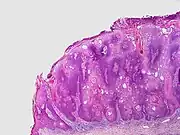
|
Differential
Dermatosis papulosa nigra (DPN) is a condition of many small, benign skin lesions on the face, a condition generally presenting on dark-skinned individuals.[19]: 638–9 DPN is extremely common, affecting up to 30% of Black people in the US.[23]
Treatment
No treatment is necessary, except for reasons of appearance.[11] Generally, lesions can be treated with electrodesiccation and curettage, or cryosurgery. When correctly performed, removal of seborrheic keratoses will not cause much scarring.[24]
Epidemiology
Seborrheic keratosis is the most common benign skin tumor. It becomes more common with age. People with darker skin are less commonly affected.[25] In one study, 100% of the people over age 50 had at least one.[26] Onset is usually in middle age, although they are common in younger people too—found in 12% of 15-year-olds to 25-year-olds—making the term "senile keratosis" a misnomer.[27]
See also
Notes
- ↑ Inverted follicular keratosis is generally thought to be a rare variant of seborrheic keratosis, but this position is not universally accepted.
- Karadag, AyseSerap; Ozlu, Emin; Uzuncakmak, TugbaKevser; Akdeniz, Necmettin; Cobanoglu, Bengu; Oman, Berkant (2016). "Inverted follicular keratosis successfully treated with imiquimod". Indian Dermatology Online Journal. 7 (3): 177–9. doi:10.4103/2229-5178.182354. ISSN 2229-5178. PMC 4886589. PMID 27294052.
References
- ↑ Paech, Volker; Schulz, Hans; Argenyi, Zsolt; Gambichler, Thilo; Altmeyer, Peter (31 July 2008). Compendium of Surface Microscopic and Dermoscopic Features. Springer Science & Business Media. p. 115. ISBN 978-3-540-78973-4. Retrieved 19 June 2025.
- ↑ 2.00 2.01 2.02 2.03 2.04 2.05 2.06 2.07 2.08 2.09 2.10 2.11 "Seborrhoeic keratoses (brown warts, basal cell papillomas, seborrheic keratosis)". DermNet®. 26 October 2023. Archived from the original on 29 July 2025. Retrieved 19 June 2025.
- ↑ 3.00 3.01 3.02 3.03 3.04 3.05 3.06 3.07 3.08 3.09 3.10 3.11 3.12 3.13 3.14 "British Association of Dermatologists". www.bad.org.uk. British Association of Dermatologists. Archived from the original on 25 March 2025. Retrieved 19 June 2025.
- ↑ 4.0 4.1 "Seborrheic keratoses: Overview". www.aad.org. AAD. Retrieved 19 June 2025.
- ↑ 5.0 5.1 5.2 "Seborrheic Keratoses - Dermatologic Disorders". MSD Manual Professional Edition. MDS. Archived from the original on 8 April 2025. Retrieved 19 June 2025.
- ↑ 6.0 6.1 6.2 Greco, MJ; Bhutta, BS (January 2025). "Seborrheic Keratosis". StatPearls. PMID 31424869.
- ↑ 7.0 7.1 7.2 7.3 7.4 "ACD A-Z of Skin - Seborrhoeic Keratoses". ACD. Australasian College of Dermatologists. Archived from the original on 2 April 2024. Retrieved 19 June 2025.
- ↑ DE, Elder; D, Massi; RA, Scolyer; R, Willemze (2018). "1. Keratinocytic/epidermal tumours". WHO Classification of Skin Tumours. Vol. 11 (4th ed.). Lyon (France): World Health Organization. pp. 23–64. ISBN 978-92-832-2440-2. Archived from the original on 2022-07-11. Retrieved 2023-07-31.
- ↑ Jackson, Scott (15 September 2022). Skin Disease and the History of Dermatology: Order out of Chaos. CRC Press. p. PT152. ISBN 978-1-000-64401-2. Retrieved 19 June 2025.
- ↑ Reutter, Jason C.; Geisinger, Kim R.; Laudadio, Jennifer (2014). "Vulvar Seborrheic Keratosis". Journal of Lower Genital Tract Disease. 18 (2): 190–4. doi:10.1097/LGT.0b013e3182952357. PMID 24556611. S2CID 26756807.
- ↑ 11.0 11.1 Moles, Freckles, Skin Tags, Benign Lentigines, and Seborrheic Keratoses Archived 2015-05-22 at the Wayback Machine from the Cleveland Clinic website
- ↑ "Heartburn". ssai-starss.com. Archived from the original on 22 February 2014. Retrieved 7 May 2018.
- ↑ Chen, Tiffany Y.; Morrison, Annie O.; Cockerell, Clay J. (2017-09-01). "Cutaneous malignancies simulating seborrheic keratoses: An underappreciated phenomenon?". Journal of Cutaneous Pathology. 44 (9): 747–748. doi:10.1111/cup.12975. ISSN 1600-0560. PMID 28589622. S2CID 11350866.
- ↑ Hanlon, Allison (2018). A Practical Guide to Skin Cancer. Springer. p. 80. ISBN 9783319749037. Archived from the original on 17 July 2021. Retrieved 22 September 2018.
- ↑ 15.0 15.1 15.2 15.3 15.4 15.5 15.6 15.7 Freedberg, et al. (2003). Fitzpatrick's Dermatology in General Medicine. (6th ed.). McGraw-Hill. ISBN 0-07-138076-0.
- ↑ Dermatosis Papulosa Nigra at eMedicine
- ↑ Stucco Keratosis at eMedicine
- ↑ 18.0 18.1 Rapini, Ronald P.; Bolognia, Jean L.; Jorizzo, Joseph L. (2007). Dermatology: 2-Volume Set. St. Louis: Mosby. p. 1665. ISBN 978-1-4160-2999-1.
- ↑ 19.0 19.1 19.2 James, William D.; Berger, Timothy G.; et al. (2006). Andrews' Diseases of the Skin: Clinical Dermatology. Saunders Elsevier. ISBN 978-0-7216-2921-6.
- ↑ Stucco Keratosis at eMedicine
- ↑ "Cutaneous Melanoacanthoma: eMedicine Dermatology". Archived from the original on 2019-07-12. Retrieved 2019-09-20.
- ↑ Tan, Kong-Bing; Tan, Sze-Hwa; Aw, Derrick Chen-Wee; Jaffar, Huma; Lim, Thiam-Chye; Lee, Shu-Jin; Lee, Yoke-Sun (2013). "Simulators of Squamous Cell Carcinoma of the Skin: Diagnostic Challenges on Small Biopsies and Clinicopathological Correlation". Journal of Skin Cancer. 2013: 1–10. doi:10.1155/2013/752864. PMC 3708441. PMID 23878739.
{{cite journal}}: CS1 maint: unflagged free DOI (link) - ↑ Grimes PE, Arora S, Minus HR, Kenney JA Jr. Dermatosis papulosa nigra. Cutis. Oct 1983;32(4):385-6, 392.
- ↑ "Seborrheic keratoses | American Academy of Dermatology". www.aad.org. American Academy of Dermatology. Archived from the original on 29 April 2021. Retrieved 22 September 2018.
- ↑ Zhang, Ru-Zhi; Zhu, Wen-Yuan (2011). "Seborrheic keratoses in five elderly patients: An appearance of raindrops and streams". Indian Journal of Dermatology. 56 (4): 432–434. doi:10.4103/0019-5154.84754. PMC 3179013. PMID 21965858.
{{cite journal}}: CS1 maint: unflagged free DOI (link) - ↑ Yeatman JM, Kilkenny M, Marks R (Sep 1997). "The prevalence of seborrhoeic keratoses in an Australian population: does exposure to sunlight play a part in their frequency?". Br J Dermatol. 137 (3): 411–4. doi:10.1111/j.1365-2133.1997.tb03748.x. PMID 9349339.
- ↑ Gill D, Dorevitch A, Marks R (Jun 2000). "The prevalence of seborrheic keratoses in people aged 15 to 30 years: is the term senile keratosis redundant?". Arch Dermatol. 136 (6): 759–62. doi:10.1001/archderm.136.6.759. PMID 10871940.
External links
| Classification | |
|---|---|
| External resources |