Amoebic liver abscess
| Amoebic liver abscess | |
|---|---|
| Other names: Amoebic hepatic abscess,Hepatic amebiasis[1] | |
  | |
| |
| Specialty | Infectious disease |
| Symptoms | Fever, right upper quadrant abdominal pain, and tenderness[2] |
| Causes | Entamoeba histolytica infection[2] |
| Risk factors | Alcoholism, immunocompromised[2] |
| Diagnostic method | Ultrasound, CT[2] |
| Differential diagnosis | Candida[2] Bacterial liver abscess Echinococcus Granulosus |
| Treatment | Metronidazole[2] |
Amoebic liver abscess is a type of liver abscess caused by amebiasis. It is the involvement of liver tissue by trophozoites of the organism Entamoeba histolytica and of its abscess due to necrosis.[2]
As to treatment we find first-line anti parasitic agents metronidazole or tinidazole are used.Amoebic liver abscess common in tropical regions with poor sanitation: Africa, India, Southeast Asia, and Latin America.[3][4][2]
Signs and symptoms
Approximately 90% of patients with E histolytica are asymptomatic.[5] The two most common manifestations of E histolytica include colitis (bloody stool with mucus, abdominal pain, and/or diarrhea), and discovery of a liver abscess on imaging.[5]Liver abscess' commonly present as right upper quadrant abdominal pain and fever, with worsening features associated with abscess rupture.[5] Some further the symptoms are:[2][3]
- Pain right hypochondrium referred to the right shoulder
- Pyrexia
- Profuse sweating and rigors
- Loss of weight
- Jaundice
- Tenderness and rigidity in right hypochondrium
- Intercostal tenderness
- Cough
Complications
As to the possible complications of Amoebic liver abscess we find the following:[2]
- Rupture into the chest, or pericardium
- Inferior vena cava thrombosis
- Hepatic vein thrombosis
Cause
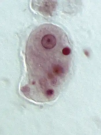
As to Amebic liver abscess it is caused by the parasite Entamoeba histolytica, which leads to condition called amebiasis or amebic dysentery[3]
Risk factors
In terms of the risk factors for Amebic liver abscess we find:[3]
- Alcoholism
- Immunosuppression
- Malnutrition
- Steroid use
Mechanism
As to the pathogenesis of Amoebic liver abscess we find that trophozoites adhere to colonic epithelial cells using Gal/GalNAc lectin.They release cysteine proteinases that degrade extracellular matrix proteins, allowing invasion into submucosal tissues.From the colon, trophozoites enter mesenteric venules, then the portal circulation, reaching the liver.Inside liver, trophozoites induce apoptosis of hepatocytes, leading to necrosis.Most commonly affects the right lobe of the liver due to its vascular supply[6][7][8][2]
Diagnosis
Diagnosis is primarily made by identifying stool ova and parasites on stool antigen testing in the presence of colitis, or E histolytica serology.[5][2][3]
- Blood ceruloplasmin
- CT scan
- Stools examination (trophozoites)
- Radiography
- Aspiration exploratory
- Medical ultrasonography
- Sigmoidoscopy
- Liver function tests
-
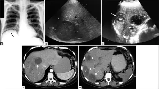 a) Amoebic liver abscess showing the elevation of the right hemi-diaphragm b)single large amoebic abscess and c) three amoebic hepatic abscesses. d) Contrasted computed tomography (CT) scan of a single abscess and e) three clear amoebic liver abscesses
 -
 Magnetic resonance cholangiopancreatography (MRC) image showing a voluminous and heterogeneous collection in the left liver lobe (amoebic abscess)
Differential Diagnosis
The DDx of Amoebic liver abscess is as follows:[2]
- Candida
- Bacterial liver abscess
- Echinococcus Granulosus
- Salmonella Typhi
Treatment
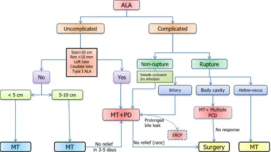
As to the management of Amoebic liver abscrss is based on Metronidazole at a dose of 500 to 750 mg orally 3 times/day; treatment should be followed by Paromomycin 500 mg 3 times/day (7 days).Due to the side effect of diarrhea Metronidazole and Paromomycin should not be given simultaneously.[2][3]
Prognosis
In terms of the prognosis of ALA we find that uncomplicated amebic liver abscesses have a good outlook. However, if an abscess breaks into the peritoneum, the mortality rate(after surgery) can be 20 to 50 percent[2]
Epidemiology
ALA accounts for up to 67 percent of liver abscess cases in endemic regions.It is endemic in tropical and subtropical regions, especially in parts of Asia, Africa, Central and South America.[3][4]
Male predominance is high, with up to 86 percent of cases occurring in men, around 40 years of age[4]
History
In terms of history we find that William Osler is credited with describing the first case of amoebic liver abscess in the United States at the end of the 19th century. Additionally, James Annesley reported the association between dysentery and amebic liver abscess in 1828.[9]
Research

Due to the difficulty of exploring host and amebic factors involved in the pathogenesis of amebic liver abscess in humans, most studies have been conducted with animal models.[10]
Histopathological findings revealed that the chronic phase of amebic liver abscess in humans corresponds to lytic or liquefactive necrosis, whereas in rodent models there is granulomatous inflammation. However, the use of animal models has provided important information on molecules and mechanisms of the host/parasite interaction in amebic liver abscess.[10]
See also
References
- ↑ "Amebic liver abscess (Concept Id: C0023886) - MedGen - NCBI". www.ncbi.nlm.nih.gov. Retrieved 20 July 2025.
- ↑ 2.00 2.01 2.02 2.03 2.04 2.05 2.06 2.07 2.08 2.09 2.10 2.11 2.12 2.13 2.14 Jackson-Akers, Jasmine Y.; Prakash, Vidhya; Oliver, Tony I. (2022). "Amebic Liver Abscess". StatPearls. StatPearls Publishing. PMID 28613582. Archived from the original on 2022-08-07. Retrieved 2022-08-05. Archived 2022-08-07 at the Wayback Machine
- ↑ 3.0 3.1 3.2 3.3 3.4 3.5 3.6 "Amebic liver abscess: MedlinePlus Medical Encyclopedia". medlineplus.gov. Archived from the original on 26 June 2025. Retrieved 24 July 2025.
- ↑ 4.0 4.1 4.2 Ghalib, Syed Muhammad Sarosh; Hashmi, Faizeaab; Zuberi, Umra Fatima; Aqeel, Sana (21 February 2025). "Epidemiology, risk factors, and clinical characteristics of amoebic liver abscess: a systematic review and meta-analysis from India". International Microbiology. doi:10.1007/s10123-025-00645-8. ISSN 1618-1905. PMID 39979548.
- ↑ 5.0 5.1 5.2 5.3 Wuerz, T; Kane, JB; Boggild, AK; Krajden, S; Keystone, JS; Fuksa, M; Kain, KC; Warren, R; Kempston, J; Anderson, J (October 2012). "A review of amoebic liver abscess for clinicians in a nonendemic setting". Canadian Journal of Gastroenterology. 26 (10): 729–33. doi:10.1155/2012/852835. PMC 3472914. PMID 23061067.
- ↑ Stanley, Samuel L. (22 March 2003). "Amoebiasis". Lancet (London, England). 361 (9362): 1025–1034. doi:10.1016/S0140-6736(03)12830-9. ISSN 0140-6736. PMID 12660071.
- ↑ 7.0 7.1 Kumar, Ramesh; Patel, Rishabh; Priyadarshi, Rajeev Nayan; Narayan, Ruchika; Maji, Tanmoy; Anand, Utpal; Soni, Jinit R. (27 March 2024). "Amebic liver abscess: An update". World Journal of Hepatology. 16 (3): 316–330. doi:10.4254/wjh.v16.i3.316. PMC 10989314. PMID 38577528.
- ↑ "Amebic Liver/Hepatic Abscesses: Background, Pathophysiology, Etiology". eMedicine. 29 March 2024. Archived from the original on 10 November 2024. Retrieved 30 July 2025.
- ↑ Arellano-Aguilar, G.; Marín-Santillán, E.; Castilla-Barajas, J. A.; Bribiesca-Juárez, M. C.; Domínguez-Carrillo, L. G. (1 October 2017). "A brief history of amoebic liver abscess with an illustrative case". Revista de Gastroenterología de México (English Edition). 82 (4): 344–348. doi:10.1016/j.rgmxen.2017.05.003. ISSN 2255-534X.
- ↑ 10.0 10.1 10.2 Campos-Rodríguez, Rafael; Gutiérrez-Meza, Manuel; Jarillo-Luna, Rosa Adriana; Drago-Serrano, María Elisa; Abarca-Rojano, Edgar; Ventura-Juárez, Javier; Cárdenas-Jaramillo, Luz María; Pacheco-Yepez, Judith (2016). "A review of the proposed role of neutrophils in rodent amebic liver abscess models". Parasite. 23: 6. doi:10.1051/parasite/2016006. ISSN 1776-1042. PMC 4754534. PMID 26880421.
External links
| Classification | |
|---|---|
| External resources |