Vorticose veins
| Vorticose veins | |
|---|---|
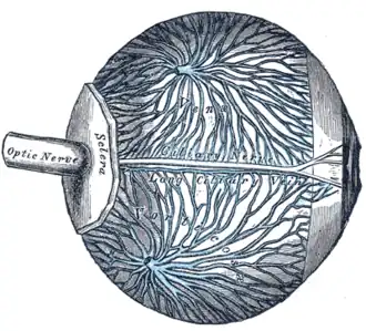 The veins of the choroid. (Venae vorticosae labeled - though difficult to see - at center.) | |
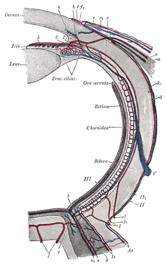 Diagram of the blood vessels of the eye, as seen in a horizontal section. ("V", at center right, is the label for the vena vorticosa) | |
| Details | |
| Drains to | Superior ophthalmic vein, and inferior ophthalmic vein |
| Artery | Short posterior ciliary arteries |
| Identifiers | |
| Latin | venae vorticosae |
| TA98 | A12.3.06.106 |
| TA2 | 4892 |
| FMA | 70880 |
| Anatomical terminology | |
The vorticose veins, commonly known as the vortex veins, are veins that drain the choroid of the eye. Usually, there are four vorticose veins in each eye, but can vary up to eight in number.[1][2] There is at least one vorticose vein per each quadrant of the eye, located at the lateral and medial sides of the superior and inferior rectus muscles. Vorticose veins drain into the superior ophthalmic vein, and inferior ophthalmic vein.[3]
Vorticose veins are an important opthalmoscopic landmark.[4]
Structure
Course and relations
Vorticose veins exit the eyeball 6 mm posterior to its equator.[3] In typical anatomy, both upper vorticose veins empty into the superior ophthalmic vein, and both lower vorticose veins drain into the inferior ophthalmic vein.[3][5]
Variation
The number of vorticose veins is known to vary from four to eight, with about 65% of the normal population having four with at least one vein in each quadrant.[3][1]
Clinical significance
Vorticose veins are an important ophthalmoscopic landmark.[4] They can be visualised in a dilated pupil using an indirect ophthalmoscope.[3]
Additional images
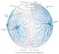 The blood-vessels of the eyeball (diagrammatic).
References
- 1 2 Verma, A; Bacci, T; Sarraf, D; Freund, KB; Sadda, SR (2021). "Vortex Vein Imaging: What Can It Tell Us?". Clinical Ophthalmology. 15: 3321–3331. doi:10.2147/OPTH.S324245. PMC 8364369. PMID 34408390.
- ↑ Kutoglu, Tunc; Yalcin, Bulent; Kocabiyik, Necdet; Ozan, Hasan (2005). "Vortex veins: Anatomic investigations on human eyes". Clinical Anatomy. 18 (4): 269–273. doi:10.1002/ca.20092. PMID 15832350. S2CID 42756249.
- 1 2 3 4 5 Remington, Lee Ann (2012). "Orbital Blood Supply". Clinical Anatomy and Physiology of the Visual System. Elsevier. pp. 202–217. doi:10.1016/b978-1-4377-1926-0.10011-6. ISBN 978-1-4377-1926-0.
- 1 2 Potter, J. W.; Vandervort, R. S.; Thallemer, J. M. (November 1984). "The clinical significance of the vortex veins". Journal of the American Optometric Association. 55 (11): 822–824. ISSN 0003-0244. PMID 6512144.
- ↑ Standring, Susan (2020). Gray's Anatomy: The Anatomical Basis of Clinical Practice (42nd ed.). New York. p. 780. ISBN 978-0-7020-7707-4. OCLC 1201341621.
{{cite book}}: CS1 maint: location missing publisher (link)